About the Tumour Bank
About the Tumour Bank - How can you simply participate in cancer research?

Contact
Secrétariat (FrontOffice)
- Tel : +32 (0)2 541 72 16
- Email : tumoroth%C3%A8que [at] bordet [dot] be (tumorothèque[at]bordet[dot]be)
Adress
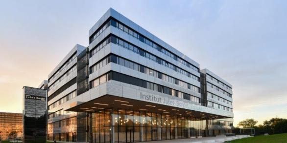
Rue Meylemeersch 90, 1070 Brussel (5th floor)
About the Tumour Bank
Our role
The Tumour Bank or Tumour Biobank at the Jules Bordet Institute is a collection of human body material samples (tumour tissue residue, blood samples) and the associated data conserved under optimal conditions for future use in research. They are the essential link between diagnosis and fundamental, translational and clinical research. Its activity is subject to strict control under the legislation.
The development of personalised medicine and immunotherapy in the field of oncology has brought the need for new biomarkers that are identifiable in tumour tissue as well as in the blood. The Tumour Bank’s role is to collect for supply to the scientific research laboratories sufficient quantities of diverse and very high quality biological samples accompanied by full clinical details to enable them to carry out their research. The aim is to understand the mechanisms of tumour development and the strategy that enables it to escape from the vigilance and immune defence of its host.
The Tumour Bank is part of the Department of Pathological Anatomy, this permitting the correct and comprehensive analysis of tumours by pathologists prior to conservation. It is a part of Belgian biobank networks – Virtual catalogue of the Cancer Register, BWB (Wallonia – Brussels biobank) and of 2 European biobanks, the BBMRI and ESBB. Through these networks the contact with the various Belgian and international research teams makes it possible to create links and share skills.
The Jules Bordet Tumour Bank received ISO9001 certification in 2012 as a guarantee of quality. The robust system put into place for the quality control of our samples enables us to support many research projects every year. The quality of the scientific publications that result is testimony to this.
The Tumour Bank in figures
- Creation : 2010
- More than 16,000 frozen tissue samples available
- Samples taken from more than 10,000 patients
- More than 65 projects using samples collected
Infrastructure projects
- Digital pathology
Digital pathology is becoming increasingly important in everyday practice, especially in research. The Jules Bordet Association financed a part of our Digital Pathology platform. The process of microscope slide digitisation enables images to be processed using computer software. A precise analysis of the various cell populations, for example the inflammatory infiltrations of solid tumours, can be carried out. These studies are transposed into clinical practice, in particular to estimate whether or not immunotherapy could benefit certain patients. The hot-spots (zones with a concentration of cell markers) are identified and analysed. . - Laser microdissection
Laser microdissection or Laser Capture/Cutting Microdissection (LCM) is a technology that makes it possible to isolate, under morphological control, cells of interest on the basis of a tissue section using a laser beam. The project supported by the Jules Bordet Association will make it possible to study the tumour heterogeneity at the cell type level.
Research projects
In addition to supplying biological material, the Tumour Bank also develops its own scientific projects.
PROJET 1
RECONNAISSANCE DES NEUTROPHILES ET MESURES DE L’EXPRESSION DE PROTÉINES CANDIDATES POUVANT AFFECTER LEURS FONCTIONS DANS LE CANCER PULMONAIRE NON À PETITES CELLULES
- Porteur de Projet : Etienne Meylan, ULB
- Collaborations : Myriam Remmelink
PROJET 2
DÉCHIFFRAGE DU RÔLE DU SÉQUENÇAGE ÉPISTRANSCRIPTOMIQUE DANS LE CANCER DU SEIN
- Porteurs de Projet : Francois Kuks, Karen Willard-Gallo, Soizic Garaud, Laboratory of Cancer Epigenetics (ULB) – Molecular Immunology Laboratory (IJB)
- Collaborations : Denis Larsimont, Ligia Craciun
PROJET 3
ROBUSTESSE DU SÉQUENÇAGE DE NOUVELLE GÉNÉRATION DE VIEUX BLOCS FFPE
- Porteurs de Projet : Ligia Craciun, Denis Larsimont, IJB
- Collaborations : Benheraoua Fatima, Spinette Alex
PROJET 4
CARACTÉRISATION DU MICROBIOTE DANS LES MÉTASTASES COLO-RECTALES : NATURE ET IMPACT SUR LE MICROENVIRONNEMENT TUMORAL IMMUNITAIRE, L’ÉVOLUTION DE LA MALADIE ET LA RÉPONSE AU TRAITEMENT
- Porteurs de Projet : Van Den Eynde Marc, Philippe Stevens, UCL
- Collaborations : Ligia Craciun, Pieter Demetter
PROJET 5
INFECTION PAR LE VIRUS DU PAPILLOMAVIRUS HUMAIN (VPH) DANS LE CANCER DE L’ŒSOPHAGE
- Porteur de Projet : Michael Herfs, GIGA-Cancer, University of Liege
- Collaborations : Pieter Demetter, Ligia Craciun
PROJET 6
ANALYSE DES SOUS-TYPES D’ASTROCYTES, DE MICROGLIES ET DE MACROPHAGES DANS LES MÉTASTASES CÉRÉBRALES
- Porteurs de Projet : Awada Ahmad, Kindt Nadège, IJB
- Collaborations : Ligia Craciun, Pieter Demetter
PROJET 7
CARACTÉRISATION MOLÉCULAIRE INTRATUMORALE DANS DIFFÉRENTS SOUS-TYPES DE CANCER DU SEIN ET LEUR MICRO-ENVIRONNEMENT À L’AIDE DES PLATEFORMES MULTI-OMIQUES
- Porteur de Projet : Christos Sotiriou, IJB
- Collaborations : Ligia Craciun, Denis Larsimont
PROJET 8
VALIDATION À HAUT DÉBIT DES LAMES DE MICROSCOPE POUR ENTÉRINER LES ALGORITHMES D’INTELLIGENCE ARTIFICIELLE QUI ANALYSENT LES SCANS NUMÉRIQUES DE CES MÊMES LAMES
- Porteurs de Projet : Ligia Craciun, Denis Larsimont, IJB
- Collaborations : Brandon Galass, FDA, Roberto Salgado
PROJET 9
ÉTUDE DE L’IMPORTANCE DE LA TROP2 EN TANT QUE MARQUEUR POTENTIEL DE RÉGÉNÉRATION ÉPITHÉLIALE DANS LES PATHOLOGIES INFLAMMATOIRES DE L’INTESTIN CHEZ L’HOMME
- Porteurs de Projet : M. Isabelle Garcia, IRIBHM, ULB Erasme
- Collaborations : Pieter Demetter, Ligia Craciun
PROJET 10
MÉCANISMES DE LA CYTOTOXICITÉ LIÉE À PDE3A DANS LES TUMEURS STROMALES GASTROINTESTINALES : ÉTUDES PRÉCLINIQUES IN VIVO ET IN VITRO
- Porteurs de Projet : Jean-Marie Vanderwinden, IRIBHM, ULB Erasme
- Collaborations : Pieter Demetter, Ligia Craciun
PROJET 11
CONTRIBUTION À L’ÉTUDE DE L’ENVIRONNEMENT IMMUN DES CANCERS DU PANCRÉAS TRAITEMENT NAÏFS ET AYANT REÇU DIFFÉRENTES TYPES DE TRAITEMENTS NÉO-ADJUVANTS
- Porteurs de Projet : Christelle Bouchart , IJB
- Collaborations : Laurine Verset, Ligia Craciun
PROJET 12
ACTIVATION DE LA PHOSPHORYLATION DU COMPLEXE CDK4 : UTILISATION POUR PRÉDIRE LA RÉACTIVITÉ TUMORALE AUX MÉDICAMENTS INHIBITEURS DU CDK4 DANS LE BUT D’ÉLARGIR LE RECOURS À CES MÉDICAMENTS DANS LES CANCERS PRIVÉS DE THÉRAPIES EFFICACES
- Porteurs de Projet : Pierre Roger, Carine Maenhaut, Thierry Berghmans, Jean-Luc Van Laethem, ULB Erasme
- Collaborations : Ligia Craciun, Denis Larsimont